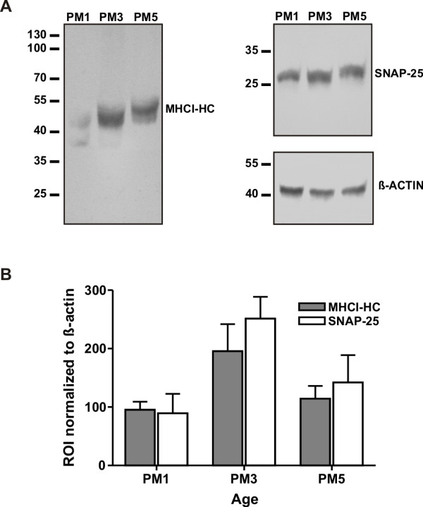
Figure 3.

MHCI-HC protein levels at different stages of visual cortex development. The antibody TP25.99 (mouse anti-human MHCI) recognized bands of appropriate size for MHCI-HC protein (MHCI-HC, A) in Western blots of proteins extracted from the marmoset visual cortex. Animals were 1, 3 and 5 months old (PM1, 3, 5) representing the main stages of synaptogenesis: initial stage, peak and rapid decline/synaptic refinement, respectively. SNAP-25 was used as a marker of synatogenesis. Data were normalized to ß-actin. Molecular weights (in kDa) are indicated on the left side. Data are from three independent experiments with N = 1 animal per stage (B).
