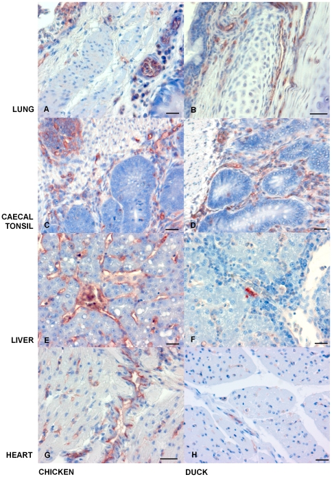
Figure 9. During H5N1 Influenza infection iNOS appears more highly expressed in chicken tissues when compared to duck tissues.
IHC for iNOS antigen staining, with left-hand panel showing chicken tissues and right-hand panels showing duck tissues. (A) Chicken lung 24 h.p.i., IHC stain (red/brown) for iNOS mainly in the submucosa. (B) Duck lung 72 h.p.i., IHC iNOS staining in the submucosa. (C) Chicken caecal tonsil 24 h.p.i., with iNOS in lymphoid follicles, the submucosa and lymphoid aggregates. (D) Duck caecal tonsil 72 h.p.i., IHC staining of iNOS mainly in the submucosa. (E) Chicken liver tissue 24 h.p.i., iNOS present in the sinusoidal endothelium. (F) Duck liver 72 h.p.i., little or no iNOS detected. (G) Chicken heart 24 h.p.i., iNOS in the myocardium and proximity to blood vessels (presumably endothelial cells). (H) Duck heart 72 h.p.i., with low levels of IHC staining for iNOS. All scale bars = 50 µm.

