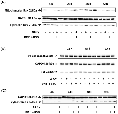
Figure 5. Translocation of the pro-apoptotic protein Bax to mitochondria and release of cytochrome c in the cytosol of irradiated GSH-depleted SQ20B cells.
SQ20B cells were treated with 100 µM DMF and 100 µM BSO for 4 h before irradiation. The drugs were then removed by washing with fresh medium. At different time post irradiation, mitochondria were isolated with standard fractionation procedure. The translocation of Bax to mitochondria (Panel A) and the release of cytochrome c in cytosol (Panel C) were measured by Western immunoblotting assay. In parallel, the activation of pro-caspase 8 and the cleavage of Bid were estimated by Western blot analysis (Panel B).

