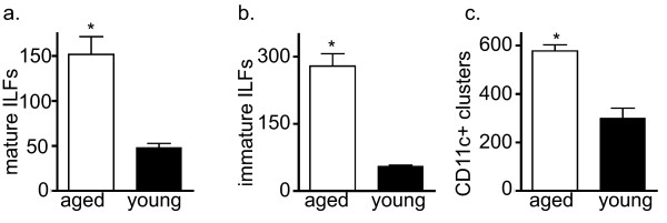
Figure 1.
ILF formation is augmented at an early stage with aging. To evaluate if the process of ILF development was altered with aging, whole mount techniques were used to identify well developed SILT containing a follicle associated epithelium indicative of mature ILFs (panel a), poorly developed or immature ILFs comprising a loose cluster of B-lymphocytes (panel b), and nascent lymphoid tissues transitioning into ILFs as identified by CD11c+ clusters (panel c) in young and aged mice. Aged mice had significantly increased numbers of mature ILFs (panel a), immature ILFs (panel b), and CD11c+ clusters (panel c), indicating that all stages of CP transitioning into ILFs are augmented with aging. n = 3 or more mice in each group. * = p < 0.05.

