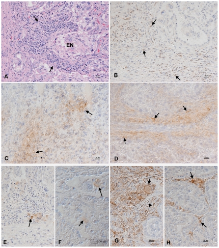
Figure 2. Histology and immunohistology in the placenta after parasite recrudescence.
Placenta after parasite recrudescence. A. Animal no. 5, 32 weeks of gestation. There is a focal area of epithelial cell necrosis (EN) and a focal lymphocyte-dominated mononuclear interstitial inflammatory infiltrate (arrows). Haematoxylin-eosin stain. B–D. Animal no. 2, 25 weeks of gestation. The inflammatory infiltrate is dominated by CD3-positive T cells (B, arrows) which are comprised of CD4+ (C) and CD8+ (D) T cells (arrows). E. Animal no. 7, 35 weeks of gestation. Macrophages, as highlighted by their expression of myeloid/histiocyte antigen (arrow) are very rare in the inflammatory infiltrates. F. Animal no. 2, 25 weeks of gestation. Neospora antigen is observed within foetal chorionic epithelial cells (top arrow) and in cell free tachyzoites (bottom arrow) in areas of epithelial cell necrosis. G. Animal no. 2, 25 weeks of gestation. MHC Class II antigen expression is seen in foetal and maternal epithelial cells (arrow) and in stromal fibroblastoid cells (arrowhead). H. Control animal, 30 weeks of gestation. There is patchy MHC Class II antigen expression by stromal fibroblastoid cells (arrows). B–H. Peroxidase anti-peroxidase method, Papanicolaou's haematoxylin counterstain.

