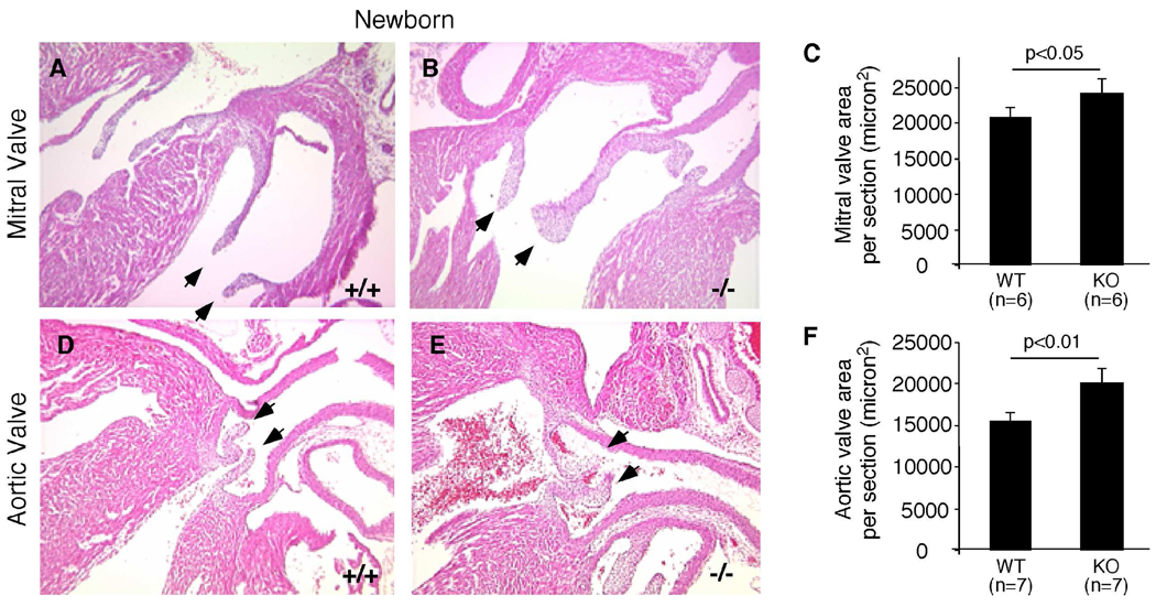
Figure 6. Valve abnormalities in ephrin-A1-deficient newborns.
Hearts from P0 wild-type (+/+) mice or knockout littermates (−/−) were processed for histological analyses. Increased thickness of mitral (B) and aortic (E) valves in (−/−) mice, compared to (+/+) controls (A,D). Arrows indicate mitral or aortic valve leaflets. (C,F) Valve areas were quantified using Image J software.

