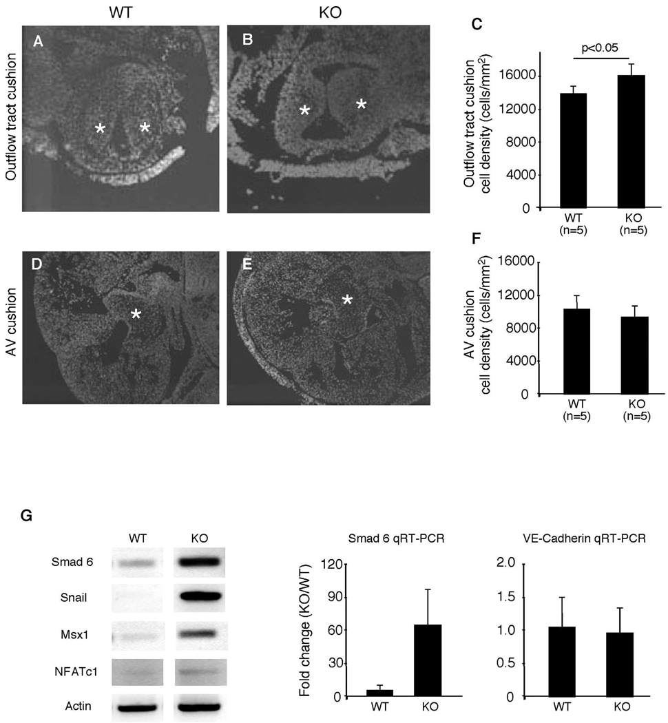
Figure 7. Reduced cellularity in Efna1−/− outflow tract cushion.
Endocardial cushions from E12.5 embryos were stained with DAPI to visualize nuclei. Outflow tract (A–C) and AV (D–F) cushion cell numbers and areas were quantified using Image J software. Cell density was calculated by dividing the number of nuclei by cushion area. Stars indicate endocardial cushion. (G) RT-PCR of mesenchymal and endocardial marker expression on mRNA from hearts of E9.5 mouse embryos. Expression of mesenchymal markers is increased in the embryonic heart. Quantitative real-time PCR of a mesenchymal and a endocardial gene, Smad6 and VE cadherin, confirm these findings.

