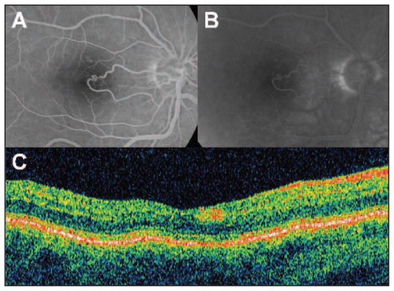
Figure 1.

This figure shows the absence of macular fluorescein leakage or other retinal pathology by standard retinal imaging. Fluorescein angiography, early (A) and late (B) phase. No leakage detected suggesting no macular edema. (C) Stratus optical coherence tomography (OCT) revealing no retinal fluid accumulation in the retina.
