Fig. 5.
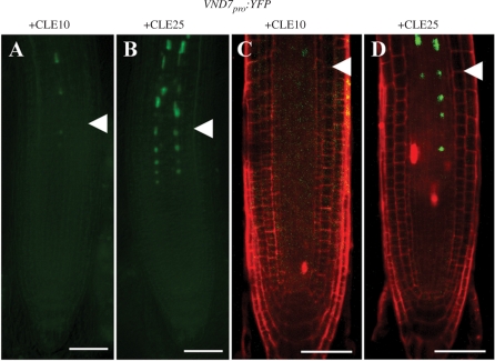
Inhibition of VND7pro:YFP expression by CLE10 peptide. (A–D) Seedlings were grown for 5 d in liquid medium with 1 μM CLE10 (A, C) or CLE25 (B, D). Fluorescence from VND7pro:YFP was detected in the non-stained primary root under a fluorescence microscope (A, B), and in the PI-stained primary root under a confocal microscope (C, D). White arrowheads represent the boundary between the RBM and the EZ. Scale bars are 50 μm.

