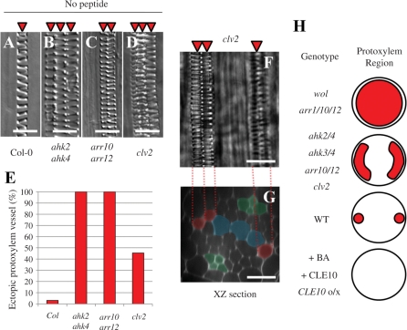
Fig. 9.
Ectopic protoxylem vessel formation in the primary root of mutants. (A–D) Protoxylem vessel formation in the WT (A), ahk2 ahk4 (B), arr10 arr12 (C) and clv2 (D). Arabidopsis roots exhibit diarch symmetry of the vascular bundle in which a protoxylem vessel file per arch is formed. However, ahk2 ahk4, arr10 arr12 and clv2 roots often have two or three protoxylem vessel files per arch. (E) The frequency of the primary root with ectopic protoxylem vessel files in the WT, ahk2 ahk4, arr10 arr12 and clv2 (N = 32–33). (F, G) Confocal images of the primary root of clv2. (F) A bright field image of the mPS–PI-stained stele. (G) An optical section of (F). Red and green indicate protoxylem vessels and phloem cells, respectively. Blue indicates undifferentiated metaxylem vessel cells. (H) Protoxylem vessel phenotypes in the primary root caused by loss-of-function and gain-of-function of CLE and cytokinin signaling. Circles show the root stele. Red indicates the area that protoxylem vessel cells occupy. CLE10o/x shows plants in which CLE10 is overexpressed. Scale bars are 25 μm.

