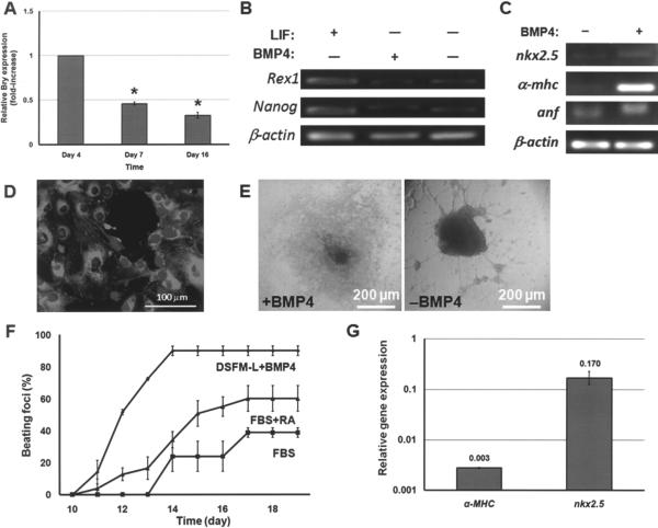
Figure 2.
(A) Mouse ESCs were cultured with DSFM-L and 10 ng/ml BMP4 for 6 days and in DSFM-L alone thereafter. The expression profile of Bry in these cells was determined by qPCR. *p < 0.001 compared to day 4 Bry expression. (B) Expression of pluripotency genes by mESCs cultured in DSFM (+LIF), DSFM-L with BMP4 (day 16), and DSFM-L without BMP4 (day 16). (C) Expression of cardiomyocyte genes after 16 days of mESC differentiation in DMSF-L with or without BMP4. (D) Cells differentiated with BMP4 exhibit α-actinin (costaining with DAPI). Day 14 cells are shown. (E) Beating clusters emerged after 10–11 days of differentiation with BMP4. However, mESCs cultured in DMSF-L alone (−BMP4) were morphologically different and no contractile foci were observed. (F) Beating curves for mESCs differentiated under different conditions. (G) Exposure to 150 ng/ml noggin (+DSFM-L + BMP4) resulted in reduced cardiac cell gene expression. Expression from cells cultured in DSFM-L + BMP4 without noggin was set to 1. Values are reported as mean ± SD.

