
Figure 3.
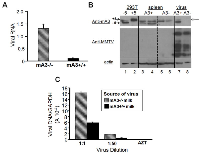
Virus isolated from mA3 −/− milk is more infectious than that isolated from mA3 +/+ milk. A) Virions were isolated from the milk of MMTV-infected mA3 −/− and +/+ mice and RNA isolated from equal amounts of milk was subjected to reverse-transcribed RT-qPCR. Error bars represent the SD of 11 mA3−/− and 9 mA3+/+ milk samples. B) Western blot analysis of APOBEC3 in tissues and virions. Lanes 1 and 2, 293T cells transfected with expression vectors containing the BL/6 delta exon 5 (−5) or BALB/c + exon 5 (+5) cDNAs; lanes 3 and 4, splenic extracts from mA3 +/+ mice; lanes 5 and 6, splenic extracts from mA3 −/− mice; lane 7 and 8, virions isolated from the mammary tissue of mA3 +/+ (A3+, lane 7) or −/− (A3−, lane 8) mice. The top panel was probed with rabbit anti-mouse APOBEC3 antisera then stripped and re-probed with goat anti-MMTV antisera (middle panel); the bottom panel is a duplicate blot of the same extracts probed with anti-β-actin. A cross-reacting protein that is present in the tissue extracts of both +/+ and −/− mice is indicated by a dotted arrow; this protein was detected in some, but not all virus preparations irrespective of the presence of APOBEC3 (not shown). C) Various dilutions of virus isolated from mA3 −/− and +/+ milk were used to infect 293-mTFR1 cells in triplicate, DNA was isolated from the infected cells and subjected to RT-qPCR for MMTV and GAPDH. The numbers presented were normalized to the amount of virion RNA as measured in A). Error bars represent SD from 3 replicate wells. p ≤ 0.001 for both dilutions.