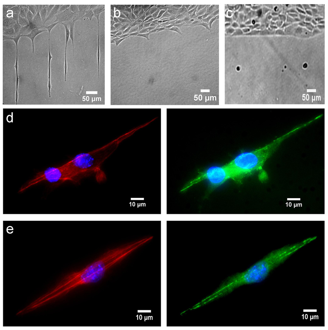
Figure 4. Cell interaction with subcellular stimuli.
(a) Cells on a 2 µm wide pattern of plasma treated polystyrene. (b) Cells on a 500 nm pattern with cells attempting to extend membrane segments onto the pattern. (c) Cells on a 100 nm wide pattern. (d) 3T3 Cells on a 1 µm wide line pattern stained for F-actin (red), vinculin (green), and nucleus (blue). (e) Cells on a 2 µm wide line pattern stained for F-actin (red), vinculin (green), and nucleus (blue). Images are representative of three experiments using identical line sizes. Cells are 3T3 fibroblasts.

