Figure 1.
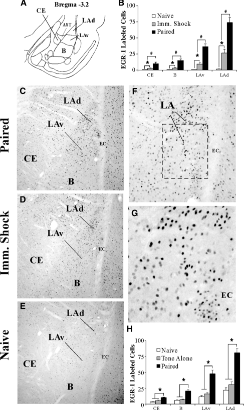
Fear conditioning regulates EGR-1 protein expression in the amygdala. (A) A representative diagram of the amygdala at ∼bregma −3.2 showing the location of the dorsal tip of the lateral nucleus (LAd), the ventral portion of the LA (LAv), the basal nucleus (B), and the central nucleus of the amygdala (CE). The amygdala-striatal transition zone (AST) is also depicted. (B) Quantification of EGR-1 labeled cells in the CE, B, LAv, and LAd of naïve (n = 3), Imm. Shock (n = 3) and Paired (n = 3) groups following fear conditioning. #P < 0.05 relative to Imm. Shock group. *P < 0.05 relative to naïve group. (C–E) Representative 10X photomicrographs of immunolabeled EGR-1 cells in a Paired, Imm. Shock, and Naïve rats, respectively. (F,G) Higher level (20X and 40X, respectively) magnifications of EGR-1 labeled cells from the Paired rat. (H) Quantification of EGR-1 labeled cells in the CE, B, LAv, and LAd of naïve (n = 5), Tone Alone (n = 7), and Paired (n = 6) groups following fear conditioning. *P < 0.05 relative to naïve and tone alone groups.

