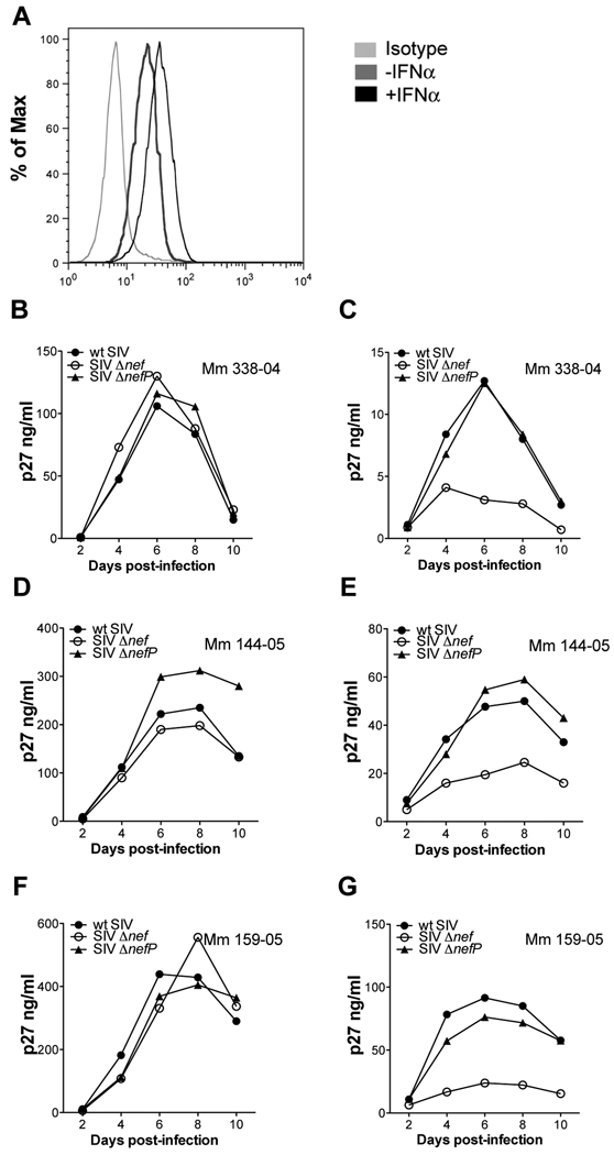
Figure 2. The compensatory changes in SIV ΔnefP enhance virus replication in interferon-treated rhesus macaque lymphocytes.
Activated primary rhesus macaque lymphocytes were infected with wild-type SIV, SIV Δnef and SIV ΔnefP. On day two post-infection, the cultures were divided and maintained in medium with or without IFNα. (A) The upregulation of tetherin on CD4+ lymphocytes was verified by flow cytometry 24 hours after treatment with IFNα. Virus replication was monitored by the accumulation of p27 in the supernatant for cultures maintained without (B, D and F), or with (C, E and G), 100 U/ml IFNα.

