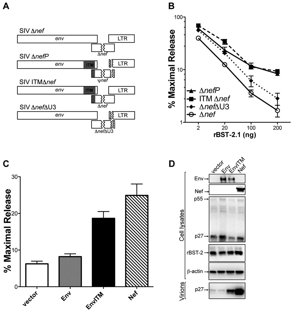
Figure 3. Amino acid substitutions in the cytoplasmic domain of gp41 account for the resistance of SIV ΔnefP to rhesus tetherin.
(A) The ITM and ΔU3 changes were engineered separately into SIV Δnef. (B) These recombinant proviruses were tested for virus release in the presence of increasing amounts of an expression construct for rhesus tetherin (rBST-2.1). (C and D) Wild-type Env, EnvITM and Nef were tested for the ability to rescue particle release for SIV ΔenvΔnef in the presence of rhesus tetherin. Virus release was measured by SIV p27 antigen-capture ELISA (C), and corroborated by comparing virion-associated p27 in cell culture supernatant to p55 Gag in cell lysates by western blot analysis (D). (B and C) Error bars represent the standard deviation (+/−) of mean percent maximal virus release.See also Figure S2.

