
Figure 1.
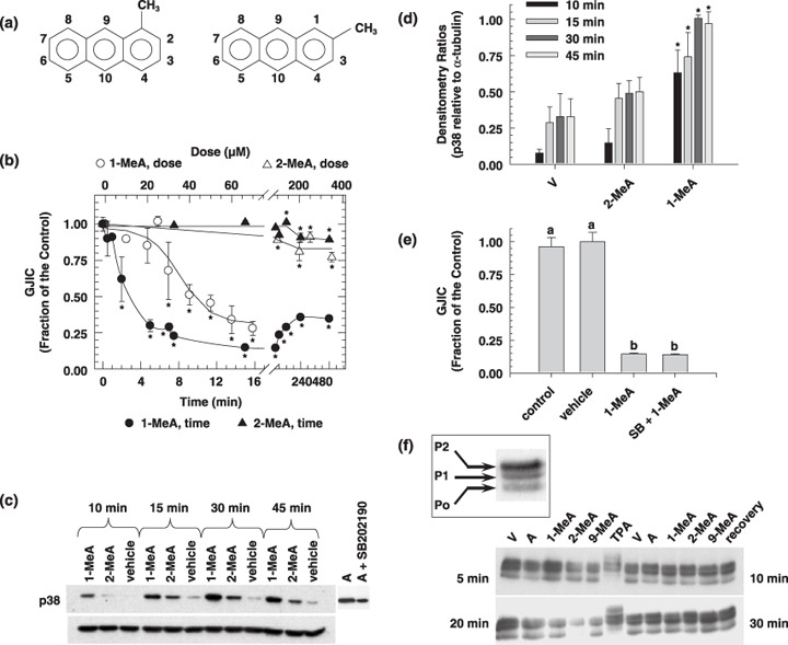
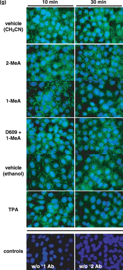
The effects of 1‐methylanthracene (1‐MeA) versus 2‐methylanthracene (2‐MeA) on gap junction biology. (a) Structures of 1‐MeA and 2‐MeA; (b) Dose and time effect of 1‐MeA versus 2‐MeA on gap junctional intercellular communication (GJIC) activity. The data for the dose–response were obtained from Rummel et al. in which WB‐F344 rat liver epithelial cell line were treated with the indicated polycyclic aromatic hydrocarbons (PAH) for 15 min. The dose for the time response data of each PAH was 75 µM, and the WB‐F344 rat liver epithelial cell line was used for this and all subsequent experiments. The scrape load‐dye transfer assay was used to assess GJIC for both the time and dose–response experiments. Average of the data (n = 3) ±standard deviation at the 95% confidence level. An anova indicated significance at P < 0.001 for 1‐MeA dose (F = 29.1), 1‐MeA time (F = 87.0), 2‐MeA dose (F = 18.2), 2‐MeA time (F = 15.9), and *indicated significance from the vehicle control using a Holm Sidak posthoc T‐test at the P = 0.05 level. (c) The effect of 1‐MeA versus 2‐MeA on the mitogen activated protein kinase, p38, as determined by Western blot analysis of p38 using phospho‐specific antibodies (top gel) and the house keeping protein α‐tubulin (bottom gel). The proteins were extracted from cells treated with 75 µM of the indicated polycyclic aromatic hydrocarbon. Anisomycin (A) at 0.5 µM and 30 min was used as a positive control. (d) Densitometry analysis of Western blots of p38 from proteins extracted from three independently treated cell culture plates, including the gel presented in (c). The values are the densitometry ratios of the p38 relative to α‐tubulin. anovas of the 10 min group (F = 23.5, P < 0.001), 15 min group (F = 9.4, P = 0.014), 30 min group (F = 34.1, P < 0.001), 45 min group (F = 31.2, P < 0.001), indicated significance between the vehicle, 2‐MeA and 1‐MeA. *Specifically indicates a difference as compared to the vehicle within each time group at P = 0.05 using a Holm‐Sidak post hoc test. (e) The effect of a p38 inhibitor on 1‐MeA‐induced inhibition of GJIC. The scrape loading‐dye transfer assay was used to measure GJIC. The dose and preincubation time for the p38 inhibitor, SB202190 (4‐(4‐Fluorophenyl)‐2‐(4‐hydroxyphenyl)‐5‐(4‐pyridyl)‐1H‐imidazole), were 20 µM and 20 min. The GJIC data are an average of the data (n = 3) ±standard deviation at the 95% confidence level. An anova indicated significance at P < 0.001, F = 285, and the different letters indicated significance using an all pair multiple comparison‐Holm Sidak posthoc T‐test at the P = 0.05 level. (f) The effect of methylanthracene isomers on the phosphorylation status of connexin43 (Cx43). A Western blot analysis using antiCx43 antibodies was used to identify the phosphorylated states of Cx43. The duration of exposure times to the various PAH and vehicle were 5, 10, 20, and 30 min as indicated in the four quadrants of the figure. The quadrants are separated by the 12‐O‐tetradecanoylphorbol‐13‐acetate (TPA) data, in which these cells were treated with TPA for 10 min. Lanes TPA = 500 ng/mL, V = vehicle (0.3% v/v acetonitrile), A = 60 µM anthracene, 1‐MeA = 60 µM 1‐methylanthracene, 2‐MeA = 60 µM 2‐methylanthracene, 9‐MeA = 60 µM 9‐methylanthracene, Recovery for top gel = 6 h recovery from cells treated with 60 µM 1‐MeA for 30 min, Recovery for bottom gel = 6 h recovery from cells treated with 60 µM 9‐MeA for 30 min. The 1st (bottom), 2nd and 3rd bands are typically designated P0, P1 and P2 as indicated in the boxed inset, and the addition of alkaline phosphatase to the protein samples results in only a P0 band, using this Western blot analysis protocol. The experiment was done in triplicate and the densitometry analyzes of the data are presented in Table 1. (g) Cytological localization of Cx43 in cells treated with 1‐MeA, 2‐MeA and TPA. The concentrations of 1‐MeA and 2‐MeA were both 70 µM, and for TPA 50 ng/mL. The vehicle was 0.3% (v/v) acetonitrile. An antiCx43 antibody was used to label this protein. The magnification of the images was 1000×. Ab, antibody; w/o, without.