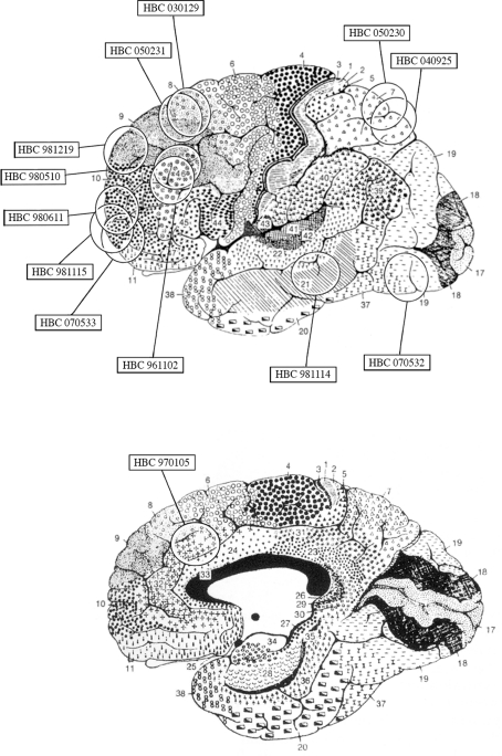
Figure 1.
Diagram based on the surgeon's estimate of cortical resections and on magnetic resonance imaging findings showing the approximate location of the cortical samples used in the present study superimposed on Brodmann's cytoarchitectonic map of the human cerebral cortex. Modified from Brodmann (1909).

