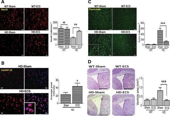
Figure 2.
ECS treatment attenuates the degeneration of striatal neurons in HD mice. (A) Representative images of NeuN-stained cells (upper micrographs; scale bar = 250 µm) and results of counts of NeuN-positive cells (lower graph) in 16-week-old WT and N171-82Q HD mice in ECS- and sham-treated groups, **P< 0.01. (B) Representative images showing DARRP-2 immunoreactivity (red; scale bar = 20 µm) and results of densitometric analysis (right graph) in 16-week-old N171-82Q HD mice in ECS- and sham-treated groups. Values are the mean and SEM (n = 9–10 mice per group). *P< 0.05. (C) Representative images of FluorJade C (FJ-C)-stained (degenerating neurons) in the striatum (micrographs at left; scale bar = 250 µm) and results of counts of FJ-C stained cells (graph) in 16-week-old WT and HD mice in ECS- and sham-treated groups. ***P< 0.001. (D) Representative images of cresyl violet-stained coronal brain sections (left) and results of measurements of the area of the lateral ventricle (graph) in 16-week-old WT and HD mice in ECS- and sham-treated groups. ***P< 0.001. Scale bar = 250 µm.

