Figure 1.
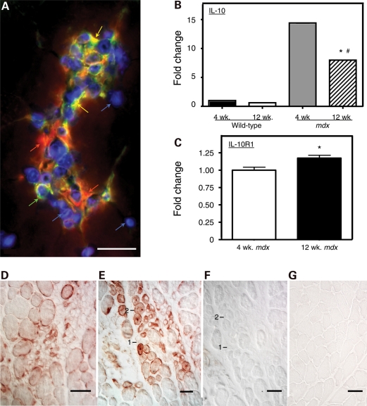
IL-10-mediated signaling between macrophages and muscle cells is enhanced in muscular dystrophy. (A) Inflamed lesions in 4-week-old mdx muscle contain M1 macrophages, M2 macrophages and satellite cells. Cross-section of quadriceps muscle was labeled with anti-F4/80 (red), which binds all macrophage phenotypes, anti-CD206 (green), which binds M2 macrophages and satellite cells, and DAPI reagent (blue), which binds DNA to show the position of nuclei. M1 macrophages are CD206−/F4/80+ (red). M2 macrophages are CD206+/F4/80+ (orange). Satellite cells are CD206+/F4/80− (green). Bar = 50 µm. (B) Quantitative, real-time PCR results for relative mRNA levels for IL-10 in quadriceps muscles of 4-week-old or 12-week-old wild-type or mdx mice. Expression levels are relative to 4-week-old wild-type muscles, for which the expression level is set at one unit. Asterisks indicate significantly different from 4-week-old wild-type quadriceps at P< 0.001. Hash symbol indicates significantly different from 4-week-old quadriceps from mice that are the same genotype at P< 0.001. Each experimental group included quadriceps from five mice. Error bars are too small to appear in the figure. (C) Quantitative, real-time PCR results for relative mRNA levels for IL-10R1 in quadriceps muscles of 4-week-old or 12-week-old mdx mice. Expression levels are relative to 4-week-old muscles, for which the expression level is set at one unit. Asterisks indicate significantly different from 4-week-old quadriceps at P< 0.05. Each experimental group included quadriceps from five mice. Bars represent sem. (D–G) Cross-sections of quadriceps muscle immunolabeled for IL-10R1 or antibody control preparations. Bars = 50 µm. (D) Section of 4-week-old mdx muscle showing an inflamed lesion containing cells expressing IL-10R1 (red). (E) Section of 4-week-old mdx muscle showing regenerative muscle fibers (labeled 1 or 2) that express IL-10R1 (red). (F) Section adjacent to the section shown in (E). The section was incubated with anti-IL-10R1 from which immunoglobins specific for IL-10R1 were depleted from the antibody solution by incubation with mouse IL-10R1 prior to labeling the section. The fibers labeled 1 or 2 are the same regenerative fibers as those labeled 1 or 2 in (E). (G) Section from 4-week-old wild-type muscle immunolabeled for IL-10R1 using treatment conditions that were identical to those used to label the mdx muscle section shown in (E).

