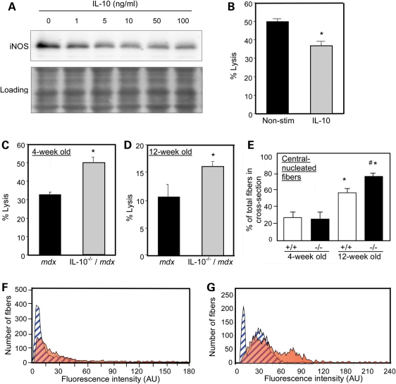
Figure 2.
IL-10 deactivates M1 macrophages and reduces muscle damage in mdx mice. (A) Macrophages isolated from 4-week-old mdx mice were stimulated with a range of IL-10 concentrations (1–100 ng/ml) and lyzed after 24 h of stimulation. Macrophage lysates were separated by SDS–PAGE and relative levels of iNOS were assayed by western blotting. The membrane used for immunoblotting was stained with Ponceau red (‘Loading’) before application of the antibody to verify equal protein loading in each lane. (B) Macrophages purified from 4-week-old mdx muscles were stimulated with IL-10 (10 ng/ml) for 24 h prior to co-culturing with C2C12 myotubes. After 24 h of co-culture, cell lysis was decreased 26% by IL-10. Asterisks indicate P< 0.05 compared with non-stimulated control. (C and D) Macrophages isolated from 4-week-old (C) or 12-week-old (D) IL-10−/−/mdx mice displayed increased cytotoxic potential compared with IL-10+/+/mdx controls (30–50% increase in IL-10−/−/mdx versus IL-10+/+/mdx). Asterisks indicates significant difference at P< 0.05 compared with IL-10+/+/mdx. (E) Quantification of the number of central-nucleated fibers in cross-sections of quadriceps muscles from 4-week-old or 12-week-old mdx mice that expressed IL-10 (+/+) or were IL-10 null mutants (−/−). Asterisk indicates significantly different from 4-week-old IL-10+/+/mdx quadriceps at P< 0.05. Hash symbol indicates significantly different from age-matched IL-10+/+/mdx mice quadriceps. Each experimental group included quadriceps from six mice. Bars represent sem. (F and G) Measurements of fluorescence intensity in muscle fibers of 4-week-old (F) and 12-week-old mice (G) indicated increases in myofiber injury in IL-10−/−/mdx mice compared with IL-10+/+/mdx controls. Blue, striped peaks represent data from IL-10+/+/mdx. Orange peaks represent data from IL-10−/−/mdx mice.

