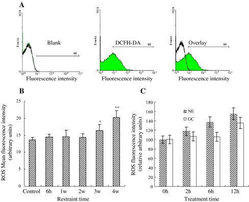
Fig. 1.
Effect of stress on ROS production in the mitochondria. a Detection of fluorescence intensity of mitochondria by dyeing with oxygen free radical fluorescent probe (DCFH-DA). b Changes of ROS content in cardiomyocytes under stress. The mitochondrial ROS content showed no significant change after 1 and 2 weeks of restraint stress, but gradually increased in the chronic stress groups to about 16, and 25% of content, after 3 and 4 weeks respectively (p < 0.05). c Changes of ROS content in cardiomyocyte treatment with GC and NE. The content increased approximately to 120, 132, and 146% in treated cardiomyocytes by NE in 2, 6, and 12 h, respectively (p < 0.05). The results of GC-treated cardiomyocytes are most similar to NE

