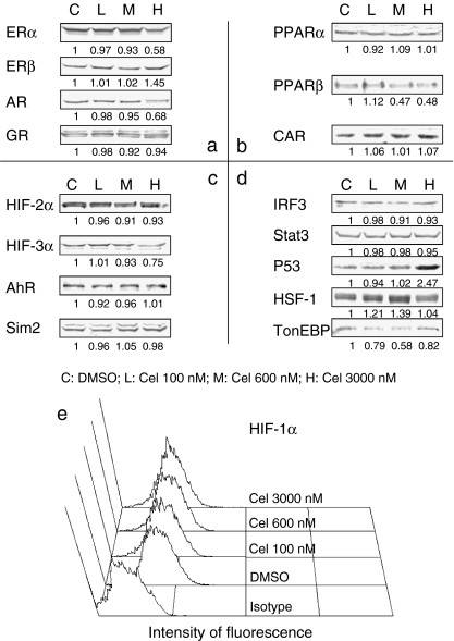
Fig. 2.
MCF-7 TFs alteration detection. MCF-7 cells were treated with different doses of celastrol for 6 h. For Western blot (WB) assay, treated cells were incubated in lysis buffer, and cell lysates were obtained by centrifugation. Equal amounts of whole cell proteins were subjected to 6–12% SDS-PAGE, as detailed in Materials and methods. For flow cytometry (FCM) assay, treated cells were washed with PBS and fixed in 100% methanol, followed by incubation in anti-HIF-1α antibodies and the corresponding secondary antibodies in conjunction with FITC, as detailed in Materials and methods. a Class I nuclear TFs detected by WB. b Class II nuclear TFs detected by WB. c Factors belonging to the PAS family except for HIF-1α detected by WB. d Other factors detected by WB. e Histogram of HIF-1α expression by FCM. Negative WB assay results are not shown. Cel celastrol. The experiment was repeated three times and the value under each band represents the mean density of the three detections

