Fig. 1.
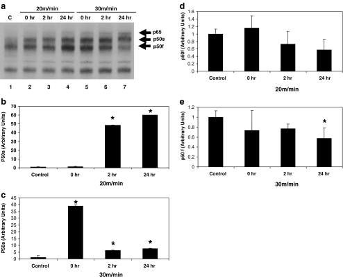
Myocardial activation of NF-κB subunit composition varies with exercise intensity and recovery from exercise. a Protein extracts were incubated with a 32P-labeled κB binding sequence and analyzed by EMSA and quantified as described in “Materials and methods”. Shown here is a portion of a representative autoradiogram. Lane 1, control (no running). Lane 2, 20 m/min, no recovery. Lane 3, 20 m/min, 2 h recover. Lane 4, 20 m/min, 24 h recovery. Lane 5, 30 m/min, no recovery. Lane 6, 30 m/min, 2 h recovery. Lane 7, 30 m/min, 24 h recovery. p65 and the fast and slow-migrating p50 NF-κB subunits are identified. b NF-κB (p50s) activation in heart extracts from animals exercised at 20 m/min. c NF-κB (p50s) activation in heart extracts from animals exercised at 30 m/min. d NF-κB (p50f) activation in heart extracts from animals exercised at 20 m/min. e NF-κB (p50f) activation in heart extracts from animals exercised at 30 m/min. Data are expressed as mean ± SD. An asterisk denotes a significant difference from control (p ≤ 0.05)

