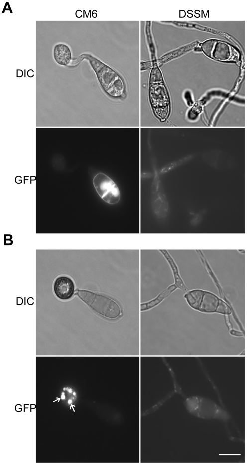
Figure 9. Expression and subcellular localization of MoMsb2-eGFP.
A. Conidia, germ tubes, and young appressoria of the MoMSB2-eGFP (CM6) and MoMSB2 ΔSP-eGFP (DSSM) transformants were examined by DIC or epifluoresence microscopy. B. In mature appressoria (24 h) of transformant CM6, GFP signals localized to small vacuole-like structures (marked with arrows). In transformant DSSM, appressorium formation was not observed and GFP signals localized in the cytoplasm. Bar = 10 µm. The same fields were examined under DIC (left) and epifluoresence microscopy (right).

