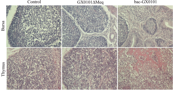
Figure 3.
Histological lesions with hematoxylin-eosin staining of bursa fabricii and thymus after inoculation with GX0101ΔMeq and bac-GX0101 at 400 × magnification. At days 28 p.i. almost all of the chickens infected with bac-GX0101 presented with obvious atrophy of bursal follicles, displayed fibrous connective tissue hyperplasia, inflammatory exudate and necrotic cells infiltrated in the follicular interstitium, vague boundary among follicular cortex and follicular medulla, rarities of follicular cortex and loss of lymphocytes in the medullary area of bursal follicles. Thymus lesions lacking structure in the cortex and medulla, necrosis and disintegration of lymphocytes were also observed in birds infected with bac-GX0101. No MD-specific lesions were observed in the control and GX0101ΔMeq-infected groups.

