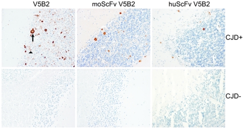
Figure 6. Immunohistochemistry of the PrPSc deposits in the cerebellum of a sCJD patient (upper three figures).
Immunolabeling was performed with whole mAb V5B2, murine scFv (moScFv) and humanized scFv (huScFv) of V5B2. The arrow marks PrPSc plaques, while the triangle marks the diffuse, synaptic PrPSc deposition. On the lower three figures immunolabeling was performed on the cerebellum of the CJD negative patient.

