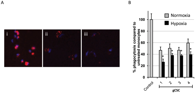
Figure 3. The ability of gCSCs to inhibit phagocytosis in monocytes is enhanced by hypoxic conditions.
A. Supernatants from gCSCs cultured in normoxic and hypoxic conditions inhibited phagocytosis of fluorescent microbeads (red) in monocytes exposed to the supernatants for 72 h, with hypoxic supernatants inhibiting phagocytosis to a greater extent than normoxic supernatants for all 4 gCSCs tested. Representative high-power fluorescent microscope images (40X) of monocytes exposed to (i) neurosphere medium alone as a positive control, (ii) normoxic gCSC supernatant, and (iii) hypoxic gCSC supernatants. Cell nuclei were stained with DAPI (blue). B. *P<0.05 indicates a significant difference between phagocytosis inhibition by hypoxic versus normoxic gCSC supernatants.

