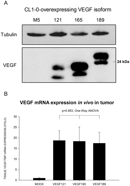
Figure 1. Western blots of VEGF isoforms proteins expression in lung cancer cells and real-time quantitative RT-PCR of VEGF isoform mRNA expression in tumor implants.
A. Western blots of VEGF isoform protein expression in cell lysate of human CL1-0 lung cancer cells transfected with single different VEGF isoform constructs. Each VEGF isoform protein comprised one glycosylated (upper) and one unglycosylated (lower) protein. Tubulin was used as an internal control. B. Quantification of VEGF mRNA expression in vivo in the tumor implants by real-time quantitative reverse transcription-PCR. The expression level of VEGF isoform was similar among CL1-0 lung cancer cell lines overexpressing one of three VEGF isoforms (p = 0.953, one-way ANOVA).

