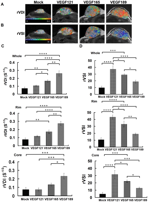
Figure 5. The rVDI and rVSI maps and values of different VEGF isofrom overexpressing tumors and the mock tumors.
In vivo rVDI and rVSI maps and quantitative curves for tumor xenografts of CL1-0 cancer cells overexpressing one of three VEGF isoforms, evaluated by SSCE-MRI. Representative high-resolution maps of the (A) rVDI and (B) rVSI in the different VEGF-overexpressing and mock tumors on day 36 after tumor implantation. In rVDI map, the color ranged from blue (0 S-1/3, lowest rVDI) to red (0.4 S-1/3, highest rVDI). In rVSI map, the color ranged from blue (0, lowest rVSI) to red (30, highest rVSI).Quantitative analysis of (C) rVDI and (D) rVSI in the whole tumor (upper), tumor rim (middle), or tumor core (lower). Differences between VEGF-overexpressing tumors and mock tumors were significant at the *p<0.05, **p<0.01, ***p<0.001, and ****p<0.0001 levels.

