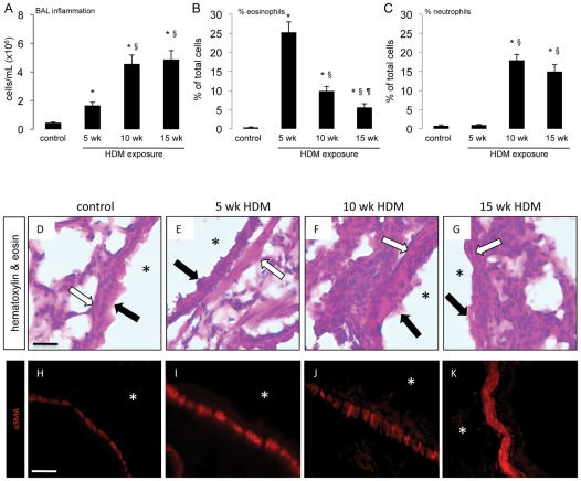
Figure 2. Prolonged respiratory HDM exposure induces inflammation.
Mice were administered sterile saline or 25 µg of HDM extract in a volume of 10 µL 5 days a week for 5, 10 or 15 consecutive weeks. (A–C) Bronchoalveolar lavage (BAL) analysis was performed to determine total inflammatory cell infiltrate (A) and to differentiate between eosinophils (B) and neutrophils (C). * p<0.05 compared to saline control animals, § p<0.05 compared to mice exposed to HDM for 5 weeks and ¶ p<0.05 compared to mice exposed to HDM for 10 weeks. Data represent mean ± SEM, n = 10–15 mice per group from two independent experiments. (D–G) Chemical staining for hematoxylin and eosin was performed on 5 µm-thick lung sections from (D) control mice exposed to saline, (E) mice exposed to HDM for 5 weeks, (F) 10 weeks or (G) 15 weeks. * indicate airway lumen, closed arrows indicate the epithelium, open arrows indicate airway smooth muscle. (H–K) Immunofluorescent staining for α-smooth muscle actin (α-SMA) was performed on 15 µm-thick lung sections from (H) control mice exposed to saline, (I) mice exposed to HDM for 5 weeks, (J) 10 weeks or (K) 15 weeks. * indicate airway lumen. Scale bar 10 µm.

