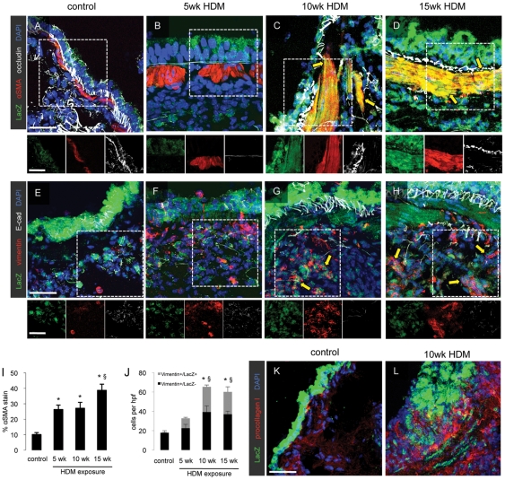
Figure 3. Prolonged respiratory HDM exposure induces epithelial-to-mesenchymal transition.
Lung sections (15 µm thick) were prepared from control mice and mice exposed to HDM for 5, 10 or 15 weeks and immunofluorescent staining for the co-expression of the LacZ reporter in airway epithelial cells with α-SMA and occludin (A–D) and with vimentin and E-cadherin (E–H) was performed. Scale bars 10 µm. Quantification of lung fibrosis was performed by morphometric analysis of lung sections stained for α-SMA (I) or LacZ and vimentin (J). * p<0.05 compared to saline control animals, § p<0.05 compared to mice exposed to HDM for 5 weeks. Data represent mean ± SEM, n = 5 mice per group. Additional lung sections were stained to detect procollagen I-producing cells in the airway wall in control mice and in mice exposed to HDM for 10 weeks (K, L).

