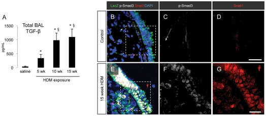
Figure 4. Activation of TGF-β signaling pathways following chronic HDM exposure.
(A) Analysis of TGF-β levels in mouse bronchoalveolar lavage (BAL) fluid in saline controls and mice exposed to HDM for 5, 10 or 15 weeks. BAL fluid was collected at the time of sacrifice and analyzed by ELISA for the expression of mouse TGF-β1. * p<0.05 compared to saline control animals, § p<0.05 compared to mice exposed to HDM for 5 weeks. Data represent mean ± SEM, n = 8 per group from two independent experiments. (B–G) Immunofluorescent staining for the expression of LacZ, p-Smad3 and Snail1 in lung sections from control mice and mice exposed to HDM for 15 weeks.

