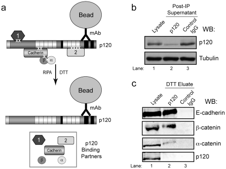
Figure 3. Elution of binding partners from p120.
(a) A schematic of the elution strategy. Following immunoprecipitation and washing of cross-linked complexes on p120 mAb beads, binding partners are released by incubation with DTT in RIPA buffer, cleaving the cross-links and releasing interacting proteins from p120. (b) A representative western blot demonstrating depletion of p120 from A431 cell lysates following immunoprecipitation with p120 mAb beads of control IgG beads. Tubulin is shown as a loading control. (c) Elution of known binding partners, but not p120, from p120 mAb beads. Whole cell lysate is shown as a control, and 10% of the DTT eluate was analyzed for E-cadherin, β-catenin, α-catenin, and p120 by Western blot.

