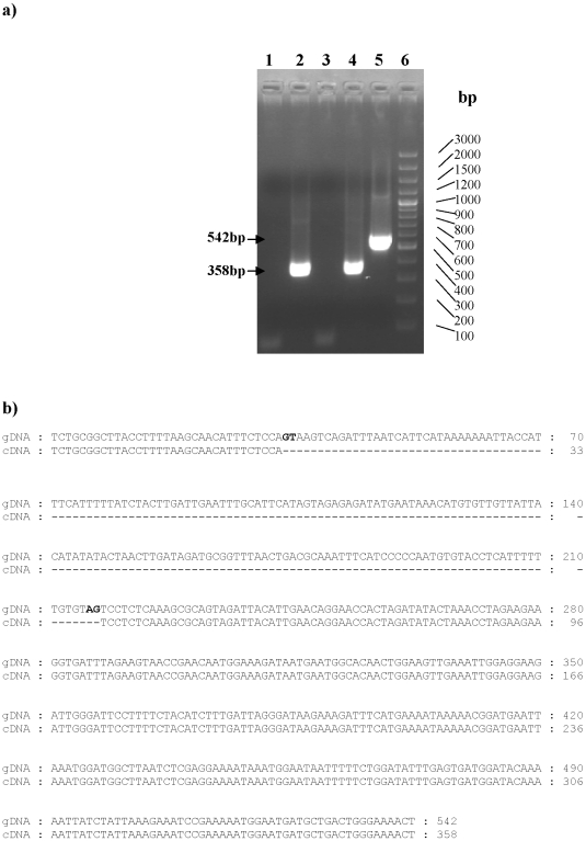
Figure 2. In vivo expression of the pvtrag33.5 gene.
a) Detection of the pvtrag33.5 gene transcript by Reverse Transcription PCR. PCR amplification of the P.vivax cDNA (Lanes 1–4) and genomic DNA (Lane 5) using pvtrag33.5 gene specific primers. RT- PCR product of pvtrag33.5 specific cDNA synthesized using oligodT (Lane 2) or random decamers (Lane 4) with gene specific primers. Reverse transcription negative control using oligodT (Lane 1) or random decamers (Lane 3) as primers for cDNA synthesis. Lane 5: PCR product of genomic DNA amplification, and Lane 6: 100 bp ladder. PCR product sizes are indicated by arrows. b) Sequence alignment of PvTRAg33.5 genomic DNA and cDNA. Splice site sequence is shown in bold letters. Dashes indicate the absence of nucleotides. Numbers on the right-hand side indicate the number of nucleotides.

