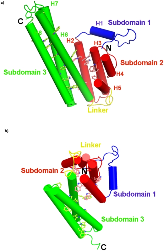
Figure 5. Model of PvTRAg33.5 structure.
a) Model of PvTRAg33.5 structure which has three subdmains: subdomain 1 (blue), subdomain 2 (red), subdomain 3 (green) and a linker (yellow) region. The helices are represented as cylinders and the coiled structure as loops. Subdomain 1 adopts a random coil structure except for a two turn helix from residues 16–25 (H1). The second subdomain has 2 long (H2: 29–54 and H3: 60–80) and 2 short helices (H4: 89–98 and H5: 104–113) connected by three loops. The subdomain 3 consists of three long helices (H6: 126–162, H7: 177–213 and H8: 217–244). The tryptophan (Trp) residues in the helices are drawn in ball-and-stick and assigned the helix color. b) The three dimensional model of PvTRAg33.5 indicating the orientation of the subdomains relative to each other at a rotation of 90° to Panel a. The linker region connecting subdomains 2 and 3 is flexible. The tryptophan (Trp) residues in the helices are drawn in ball-and-stick and assigned the helix color.

