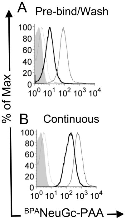
Figure 2. BPANeuGc-PAA internalization in mouse primary B cells.
Primary murine B cells were incubated with biotinylated BPANeuGc-PAA and streptavidin-PE at 4 °C, and either (A) washed to remove unbound BPANeuGc-PAA prior to warming to 37 °C; or (B) not washed before warming to 37 °C. Shown is cell-associated BPANeuGc-PAA for acid washed cells following the initial 4 °C incubation (thin grey line); total cell associated BPANeuGc-PAA after warming to 37°C (thin black line), and internalized BPANeuGc-PAA resistant to an acid wash (thick black line). Filled grey trace represents background fluorescence of the cells with no BPANeuGc-PAA.

