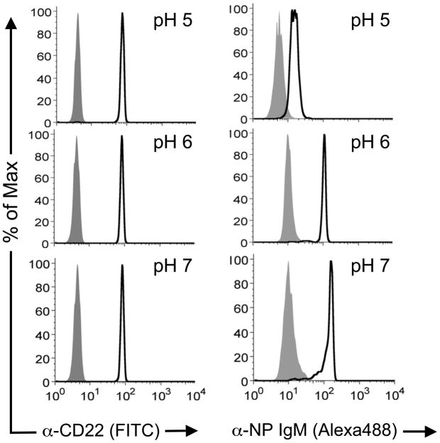
Figure 4. Differential pH dependence for αCD22 binding and BPCNeuAc-NP mediated αNP binding CD22.
CD22-Fc loaded Protein A magnetic beads were incubated with αCD22 or αNP +/− BPCNeuAc-NP at pH values 5–7 at 37 °C for three hours, then washed and analyzed by flow cytometry. Black traces are αCD22 (left panels) and αNP with BPCNeuAc-NP (right panel). Grey filled traces are isotype control (left panel) and αNP alone (right panel).

