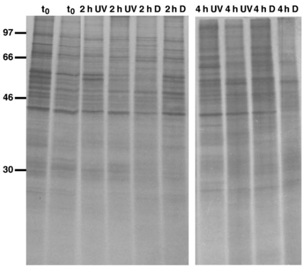
Fig. 3.

De novo protein synthesis pattern of A. tonsa after exposure to low solar radiation intensities (UV) or when kept in the dark (D) for 2 h and 4 h, respectively; t0: time zero group (17 October 2003). There was no effect of UVR on protein synthesis pattern at low solar radiation levels. Following exposure copepods were radiolabelled (35S-labelled methionine/cysteine) for 5 h at ambient seawater temperature, and proteins were separated on 12% SDS-polyacrylamide gels, which were dried and exposed to X-ray films; ~77,000 c.p.m. (left panel) and ~52,000 c.p.m. (right panel) per individual lane were loaded; each lane represents a single copepod, and individual samples were run in duplicate. The positions of 14C-labelled protein molecular mass markers are indicated in kDa on the left.
