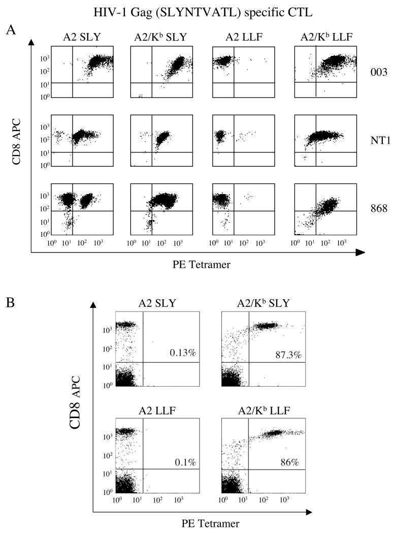
Figure 1. The exquisite specificity of pMHCI tetramer staining is lost when the strength of the pMHCI/CD8 interaction is increased by ~15-fold.
A. The 003 or NT1 CTL clones (105 cells) or the 868 CTL line (2.5×10⁵ cells), all specific for HIV-1 p17 Gag77-85, were stained with 1μg of the PE-conjugated tetramers A2 SLYNTVATL, A2/Kb SLYNTVATL, A2 LLFGYPVYV or A2/Kb LLFGYPVYV in 20μl PBS for 20 minutes at 37°C. Cells were then stained with APC-conjugated anti-CD8 and 7-AAD for 30 minutes on ice, washed twice and resuspended in PBS. Data were acquired using a FACSCalibur flow cytometer and analyzed with CellQuest software. B. 2.5×10⁵ PBMC were suspended in 250 μl FACS buffer (2% FCS/PBS), then stained with 1μg of the PE-conjugated tetramers A2 SLYNTVATL, A2/Kb SLYNTVATL, A2 LLFGYPVYV or A2/Kb LLFGYPVYV for 20 minutes at 37°C. Each sample was subsequently stained with APC-conjugated anti-CD8, PerCP-conjugated anti-CD3 and 7-AAD for 30 minutes on ice, washed twice and resuspended in FACS buffer. Data were acquired using a FACSCalibur flow cytometer and analysed with CellQuest software by gating on the live CD3+ population. The values shown represent the percent of CD3+CD8+ cells that stain with the indicated tetramer.

