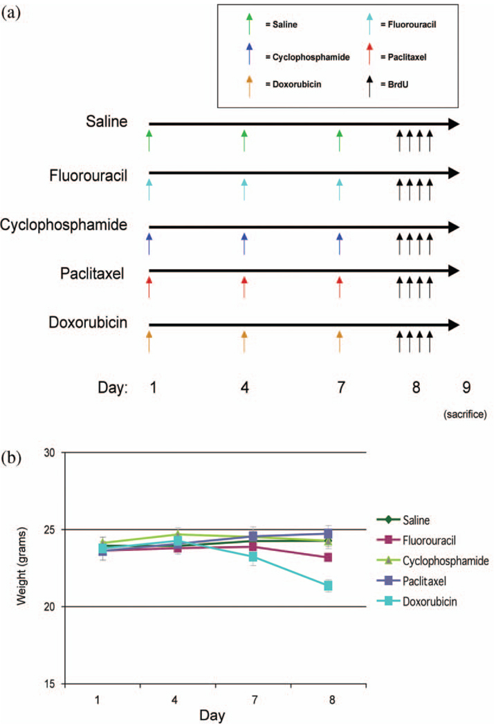
Figure 1.
Paradigms used to investigate differences in neural cell proliferation in the neurogenic dentate gyrus following administration of chemotherapeutic agents readily able to cross the BBB and those agents not readily able to cross the BBB. (a) Six–eight week old C57BL/6 mice were injected intraperitoneally with saline (control), fluorouracil, cyclophosphamide, placlitaxel, or doxorubicin on days 1, 4, and 7 (n = 6–8/group). On day 8, mice were injected intraperitoneally 4 times with BrdU to label newly divided cells. On day 9, (20–24 hr after the first BrdU injection), mice were sacrificed by transcardiac perfusion, and brains were isolated and processed for immunohistochemical analysis. (b) Mouse weights were recorded each morning prior to chemotherapy (day 1, 4, and 7) and the morning after chemotherapy (day 8). Significant differences were determined by repeated measures ANOVA considering a p < .05 for drug by day effects to be considered statistically significant.

