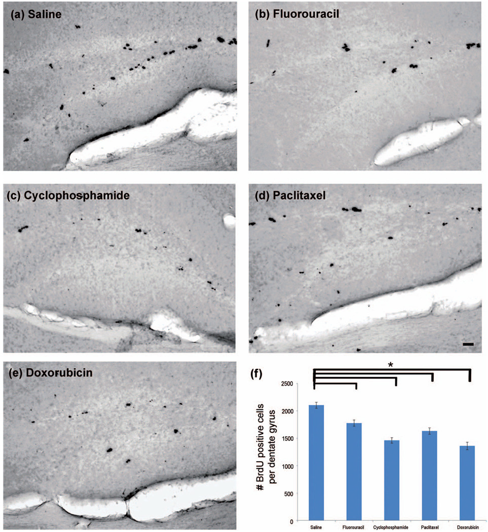
Figure 2.
Chemotherapeutic agents readily able to cross the BBB and those not readily able to cross the BBB result in a similar reduction in the number of newly divided neural cells in the neurogenic dentate gyrus. Brains were sectioned coronally at 50 µm, and immunohistochemistry was performed on every sixth section of the dentate gyrus for a total of approximately 8 free-floating sections per animal (n = 6–8/group) using the DAB method. (a–e) Representative photomicrographs display BrdU positive cells mostly within the SGZ of the dentate gyrus for each experimental group. Images of the dentate gyrus were captured, and positive cells within the SGZ, GCL and hilus were enumerated for each mouse. (f) Data are expressed as the averaged number of BrdU positive cells per dentate gyrus for each group. One-way ANOVA was performed with Dunnett’s test using saline as the control. The “*” indicates a significant (p < .05) difference compared with the saline control. The scale bar represents 50 µm.

