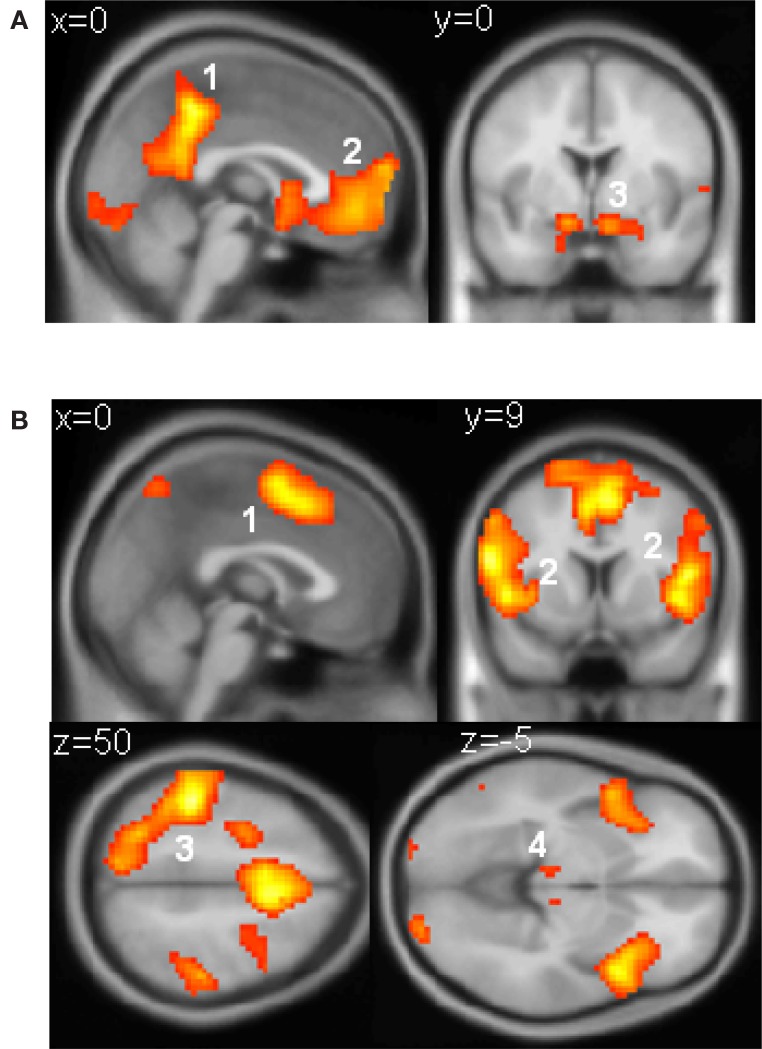
Figure S1.
(A) Voxels responding to reward. Responses that scaled with reward obtained (−5, −3, −1, 1, 3, or 5 points) were observed in (1) the posterior cingulate cortex (peak: −3, −45, 48, T(20) = 12.84), (2) the ventromedial prefrontal cortex (peak: 0, 45, −9, T = 7.39), and (3) the ventral striatum (peak right: 12, 6, −15, T(20) = 8.26; peak left: −12, 6, −15, T(20) = 7.13), as well as in the visual cuneus and lateral OFC. (B) Voxels responding to parametrically with increasing reaction time. These were found in (1) the SMA/preSMA (peak: −3, 6, 51, T(20) = 9.09), (2) the anterior insular cortex (peak right: 36, 21, 3, T(20) = 10.1; peak left: −48, 12, 0, T(20) = 9.45), (3) the parietal (peak: −45, −33, 54, T(20) = 12.98) and premotor (peak: −24, −6, 60; T(20) = 10.72) cortices predominantly on the left, and the (4) the thalamus (peak left: −9, −18, 6, T(20) = 5.84; peak right: 9, −18, 9, T(20) = 7.2) as well as in more dorsal prefrontal regions. All voxels reported in this figure survive an uncorrected threshold of at least p < 1 × 10⁻⁵).

