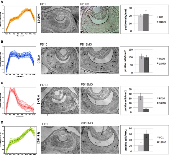
Figure 7.
Temporal expression change of VR genes confirmed by in situ hybridization. One gene was selected from four main patterns shown in Figure 5 for in situ hybridization. The number of VR expressing vomeronasal sensory neurons at different developmental time points was counted with representative slides. In situ hybridization was performed in coronal sections of mouse VNO using digoxigenin-labeled antisense RNA probes. For each pattern from (A) to (D), the overall time course expression patterns were shown, followed by two representative sections with positive label cells at two ages, then summarized the quantification of the positive labeled cells for each VR gene at each age. For each bar, three replicate mice were used. Section scale bar = 100 μm. (A,B) p > 0.1. (C,D) p < 0.05.

