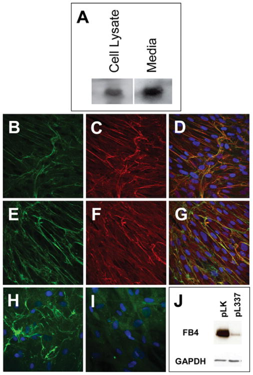
Figure 3. Characterization of fibulin-4 produced in HFF cells.
(A) Western-blot analysis of fibulin-4 in HFF cell lysate and cultured medium. (B–G) Confocal images of passage-10 HFF cultures immunostained with antibodies against human fibulin-4 (B, E), human elastin (C) and human fibrillin-1 (F). Co-localization of fibulin-4 with elastin and fibrillin-1 are shown in the merged images, (D) and (G) respectively. (H, I) Fluorescent images of HFF cultures infected with pLK virus (H) or pL337 virus (I) and immunostained with fibulin-4 antibody. (J) Western-blot analysis of fibulin-4 (FB4) in HFF cells infected with the control pLK lentivirus or the fibulin-4 shRNA-producing lentivirus pL337. Note that pL337 infection led to the abrogation of fibulin-4 staining in the ECM (I) and a significant decrease in the amount of fibulin-4 in the HFF cells (J).

