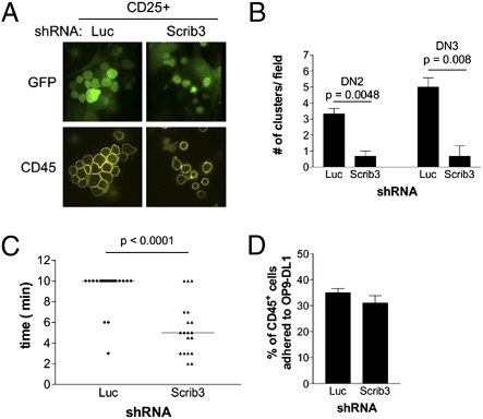
Fig. 3.
T-cell–T-cell clustering is limited in the absence of Scribble. (A) Cluster formation among FACS-purified GFP+CD25+ cells adhered to OP9-DL1 cells. (B) Quantification of clusters formed by FACS-sorted DN2 and DN3 cells. The number of clusters within the center field of view is presented. Data represent the mean ± SEM of cultures grown in triplicate, repeated in three independent experiments. (C) Motility of Luc KD and Scrib3 KD immature T cells. Data show the time period during which a cluster remained intact, with each solid circle (Luc KD) or triangle (Scrib3 KD) representing an imaged cluster. The solid bar represents the mean time of cluster maintenance. (D) Scrib3 and Luc KD cells, previously sorted as CD44highCD25+, were plated on OP9-DL1 cells and allowed to adhere for 16 h. Suspension and adherent cells were counted by flow cytometry after staining for CD45. The average percentage of adherent CD45+ cells from all experiments is shown.

