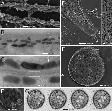
Fig. 2.
TEM and SEM micrographs of Mbav magnetosome chains (see Fig. S3 for an enlarged version at higher resolution). (A) SEM microcraph of a cryofractured cell (after chemical fixation) showing two bundles of magnetosome strands. (B and C) TEM ultrathin sections of high-pressure frozen and freeze-substituted cells showing strands of magnetosomes aligned parallel to a tubular filamentous structure (asterisk, framed area; MM, magnetosome membrane). (D and E) Cryo-SEM (frozen hydrated) of tangential (D) and cross-fractured (E) cells of Mbav (rectangular frame, magnetosomes aligned along MF; solid circles, magnetosomes crystals; dotted circle, empty MM vesicles). (F and G) SEM of focused ion beam (FIB) sections (F), and high-pressure frozen and freeze-substituted (G) Mbav cells. Circles indicate several rosette-like magnetosome bundles. Different micrographs in G represent selected sections from FIB-milling series (every 10th section is shown from left to right). Each section has a thickness of 8 nm.

