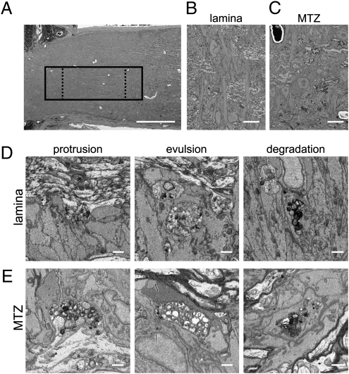
Fig. 3.
ONH astrocytes internalize large axonal evulsions. (A) Low-power view of the ONH shows the location of one of two volumes acquired (solid box) and the regions used for counting granule accumulations in the lamina and MTZ (dashed boxes). (B) Low-power scanning EM view shows transverse astrocytes at the lamina. (C) Low-power scanning EM view of MTZ shows that astrocyte somata are also transversely oriented at the onset of myelination. In both the lamina (D) and the MTZ (E), granule accumulations exist as swellings and protrusions in axons (Left), as large evulsions completely separated from axons and completely ensheathed by astrocytes (Center), and as accumulations within the cytoplasm of astrocytes (Right). (Scale bars: A, 100 μm; B and C, 10 μm; D and E, 1 μm.)

