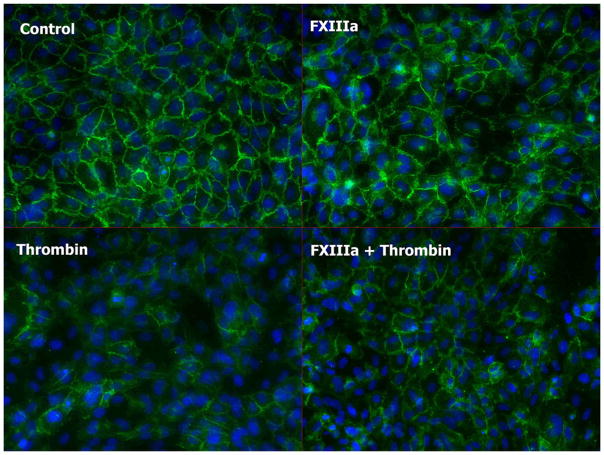
Fig. 7. Immunofluorescent staining of HUVECs treated with the following agents: thrombin, combination of thrombin and rFXIIIa, rFXIII, or albumin (control).
Cells are fixed in 4% paraformaldehyde and stained with anti–VE-cadherin antibody (Cell Signaling, Beverly, Mass), Alexa-Fluor488–conjugated secondary antibody (Invitrogen, Carlsbad, Calif), and DAPI (blue) at 60 min after treatment. Disruption of adherens junctions by thrombin is seen by the decrease in green signal intensity. Addition of rFXIIIa reduces HUVEC alterations.

