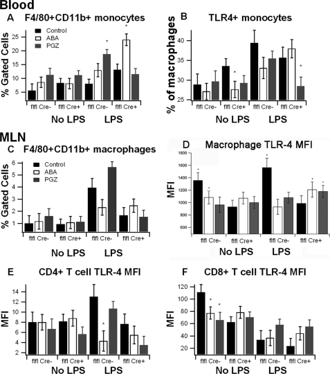
FIGURE 4.
Effect of ABA on TLR-4 expression in blood and MLN-derived immune cells in mice challenged with LPS. PPAR γ flfl; Cre− (flfl-Cre−) and PPAR γ flfl; MMTV Cre+ mice (flfl-Cre+), which lack PPAR γ in hematopoietic cells, were fed a control diet or diets supplemented with ABA or PGZ. Mice were injected with LPS (375 μg/kg) and euthanized after 6 h. Flow cytometry was performed on cells derived from blood and MLN to assess immune cell subsets affected by diet. Data are presented as means ± S.E. Data points with an asterisk indicate a significant difference from the respective control diet (p < 0.05). Results are presented as means ± S.E. of groups of 10 mice.

